
A nervous system is the most complex system of our body. It controls all types of activities of the body and it works faster than other systems of the body. The nervous system receives signals or stimuli in our body, whether it is from the external part of the body or from the internal part. These stimuli are analyzed and collected by the nervous system and produce coordinated responses, which stimulate or reduce the stimuli of the muscles, glands, and other cells of the body. The body functions smoothly through this amazing response
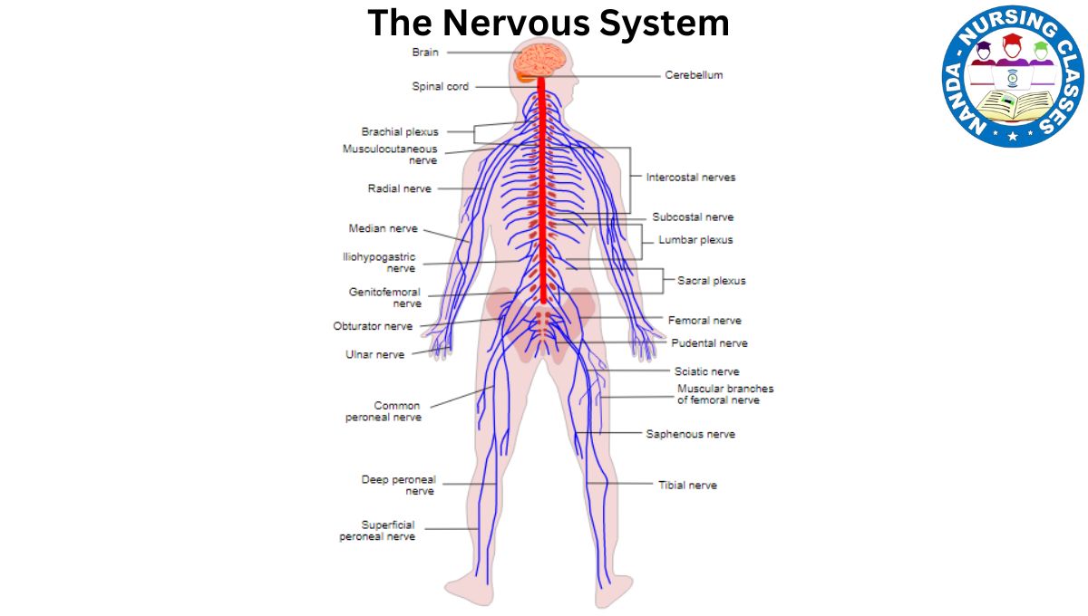
The structure of the nervous system is made up of Neuron cells and Neuroglia cells. These Neuron cells and Neuroglia cells form the nervous tissue, and the nervous system’s organs are formed from the nervous tissue. On the basis of the functionality of the nervous tissue and the organs of the nervous system, they have been classified into 2 parts. The nervous system is divided into two main branches: the central nervous system and the peripheral nervous system. The central nervous system consists of the Brain
Neuron
A neuron is the fundamental unit of the nervous system, specialized for transmitting information throughout the body. Its structure is uniquely designed to facilitate communication between different parts of the body and the brain. While neurons vary in shape and size depending on their function and location, they share a common structural organization. Understanding the anatomy and physiology of neurons is essential for nursing students, as it lays the groundwork for comprehending how the nervous system functions and how various conditions can affect it. Here’s a detailed overview of neuron structure and function.Below is a detailed explanation of the key components of a neuron:
Anatomy of Neurons
Neurons are specialized cells designed for communication within the nervous system. They consist of several key components:
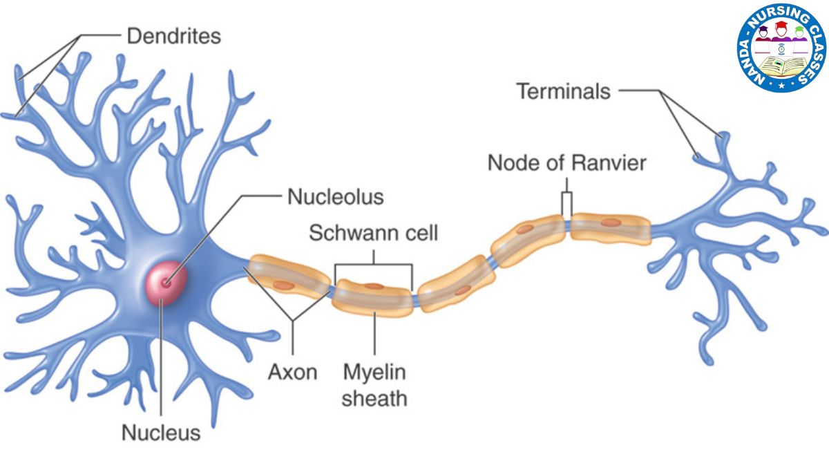
Cell Body (Soma)
The cell body is the central part of the neuron, containing the nucleus, which houses the cell’s genetic material. This area is responsible for the metabolic activities of the neuron, including protein synthesis and energy production. The cell body integrates signals received from the dendrites and determines whether to generate an action potential.
Dendrites
Dendrites are short, branching extensions that emerge from the cell body. Their primary role is to receive incoming signals from other neurons or sensory receptors. The extensive branching increases the surface area available for receiving signals, allowing a neuron to connect with multiple other neurons simultaneously.
Axon
The axon is a long, slender projection that transmits electrical impulses away from the cell body. It can vary in length, with some axons extending over a meter in the case of motor neurons. Key features of the axon include:
- Axon Hillock: The region where the axon begins, crucial for initiating action potentials.
- Myelin Sheath: A fatty layer that insulates the axon, produced by Schwann cells in the peripheral nervous system and oligodendrocytes in the central nervous system. This sheath increases the speed of electrical signal transmission.
- Nodes of Ranvier: Gaps in the myelin sheath that facilitate rapid signal conduction through a process called saltatory conduction, where the action potential jumps from node to node.
Axon Terminals
At the end of the axon, the axon terminals form synapses with other neurons or target cells (like muscles or glands). These terminals contain synaptic vesicles filled with neurotransmitters, which are released into the synaptic cleft to communicate with the next cell.
Physiology of Neurons
The physiology of neurons involves the processes by which they transmit signals. Here are the key physiological aspects:
- Resting Potential:
Neurons maintain a resting potential, which is a stable, negative charge inside the cell compared to the outside. This is primarily due to the distribution of ions, particularly sodium (Na⁺) and potassium (K⁺), across the neuron’s membrane. The sodium-potassium pump actively transports Na⁺ out of the cell and K⁺ into the cell, contributing to this resting state. - Action Potential:
When a neuron is stimulated, it can generate an action potential, which is a rapid change in membrane potential. This occurs when sodium channels open, allowing Na⁺ ions to flood into the cell, making the inside more positive. This electrical impulse travels along the axon to the axon terminals. - Synaptic Transmission:
Upon reaching the axon terminals, the action potential triggers the release of neurotransmitters into the synaptic cleft. These chemicals bind to receptors on the postsynaptic neuron, leading to either excitation or inhibition of that neuron. This process is crucial for communication within the nervous system. - Refractory Period:
After an action potential, the neuron enters a refractory period during which it cannot fire another action potential. This ensures that signals travel in one direction and prevents excessive stimulation of the neuron.
Neuroglia
Neuroglia, also known as glial cells, are non-neuronal cells in the nervous system that provide essential support, protection, and maintenance for neurons. Unlike neurons, which are responsible for transmitting electrical signals, neuroglia play a supportive role that ensures the proper functioning of the nervous system. Understanding the anatomy and physiology of neuroglia is crucial for nursing students, as these cells are involved in many neurological processes and disorders.
Anatomy of Neuroglia
Neuroglia are smaller than neurons but far more numerous, outnumbering neurons by about 10 to 1. They are found in both the central nervous system (CNS) and the peripheral nervous system (PNS). The types of neuroglia differ between these two systems:
- Neuroglia in the Central Nervous System (CNS):
-
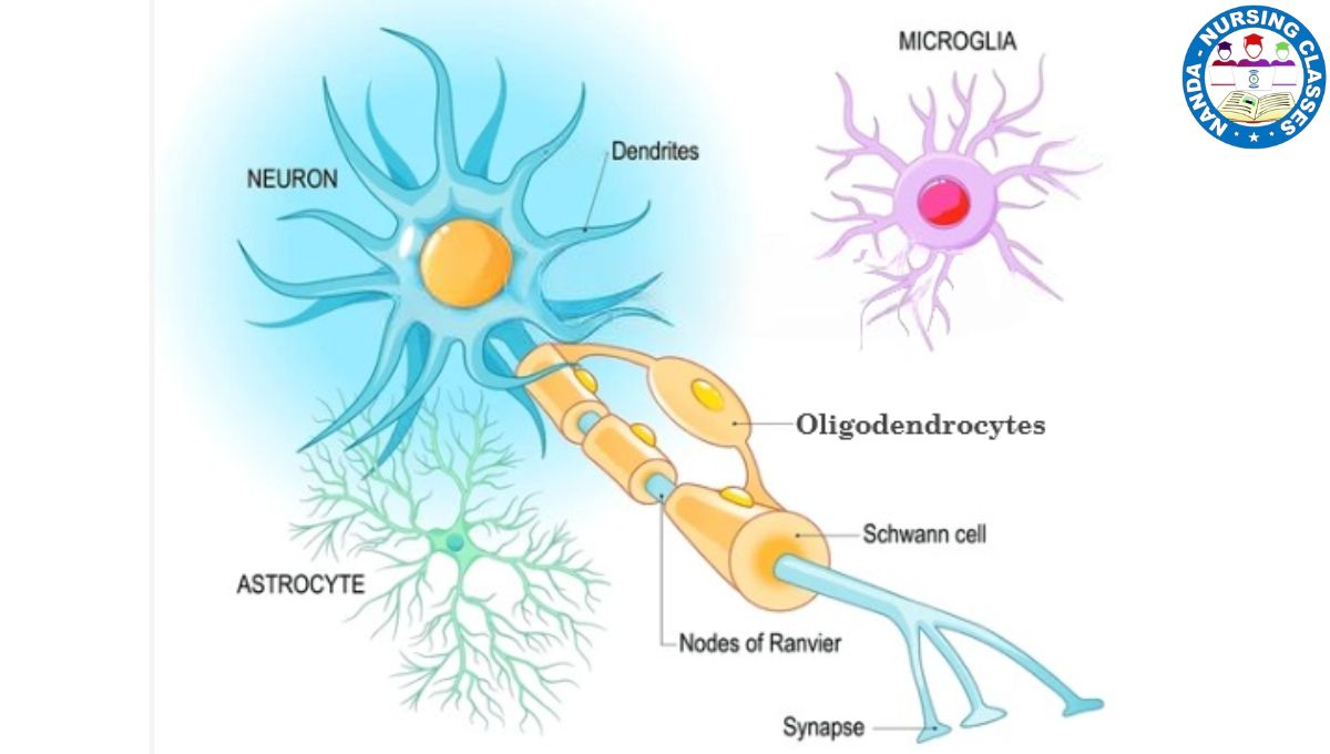
- Astrocytes:
Astrocytes are star-shaped cells that are the most abundant type of glial cells in the CNS. They have long processes that extend to neurons and blood vessels. Their structure allows them to form a supportive framework for neurons and maintain the blood-brain barrier, which protects the brain from harmful substances in the bloodstream. - Oligodendrocytes:
These cells are responsible for forming the myelin sheath around axons in the CNS. Myelin is a fatty substance that insulates axons, allowing electrical signals to travel faster. Each oligodendrocyte can myelinate multiple axons. - Microglia:
Microglia are small, mobile cells that act as the immune defense of the CNS. They constantly monitor the environment for signs of infection or injury and remove debris, dead cells, and pathogens through phagocytosis. - Ependymal Cells:
These cells line the ventricles of the brain and the central canal of the spinal cord. They are involved in producing and circulating cerebrospinal fluid (CSF), which cushions the brain and spinal cord and helps maintain a stable environment.
- Astrocytes:
- Neuroglia in the Peripheral Nervous System (PNS):
- Schwann Cells:
Schwann cells perform a similar function to oligodendrocytes but in the PNS. They form the myelin sheath around peripheral axons and play a critical role in nerve regeneration after injury. - Satellite Cells:
These cells surround the cell bodies of neurons in the PNS, particularly in ganglia. They provide structural support and help regulate the exchange of nutrients and waste products between neurons and their environment.
- Schwann Cells:
Physiology of Neuroglia
The physiological roles of neuroglia are diverse and essential for maintaining the health and functionality of the nervous system. Below are the key functions of neuroglia:
- Support and Protection:
Neuroglia provide structural support to neurons, ensuring that they remain in the correct position and are protected from mechanical damage. Astrocytes, in particular, form a supportive framework for neurons and blood vessels. - Myelination:
Oligodendrocytes in the CNS and Schwann cells in the PNS produce the myelin sheath, which insulates axons and increases the speed of electrical signal transmission. This process, known as myelination, is critical for efficient communication within the nervous system. - Immune Defense:
Microglia act as the immune cells of the CNS. They detect and respond to infections, inflammation, and injury by engulfing pathogens and debris. This function is particularly important in conditions like neurodegenerative diseases, where inflammation plays a significant role. - Regulation of the Extracellular Environment:
Astrocytes help maintain the chemical balance in the extracellular environment by regulating ion concentrations, removing excess neurotransmitters, and providing nutrients to neurons. This ensures that neurons can function optimally. - Cerebrospinal Fluid Production and Circulation:
Ependymal cells produce and circulate cerebrospinal fluid (CSF), which cushions the brain and spinal cord, removes waste products, and provides nutrients. - Nerve Regeneration:
Schwann cells in the PNS play a vital role in nerve repair and regeneration. After an injury, they guide the regrowth of axons and help restore nerve function.
Anatomy and Physiology of the Central Nervous System
The Central Nervous System (CNS) is a crucial part of human anatomy, comprising the brain and spinal cord. It serves as the primary control center for processing information, coordinating movements, and regulating bodily functions. For nursing students, understanding the anatomy and physiology of the CNS is essential for effective patient care and management of neurological conditions.
Anatomy of the Central Nervous System
The Brain
The brain is the most complex organ in the body and is responsible for interpreting sensory information, controlling motor functions, and enabling cognition and emotions. It consists of several key parts:
- Cerebrum: The largest portion of the brain, divided into two hemispheres (right and left) and four lobes:
-
-
-
- Frontal Lobe: Involved in reasoning, problem-solving, and voluntary movement.
- Parietal Lobe: Processes sensory information, such as touch, temperature, and pain.
- Temporal Lobe: Responsible for auditory processing and memory.
- Occipital Lobe: Primarily responsible for visual processing.
-
-
- Cerebellum: Located at the back of the brain, it coordinates movement, balance, and posture.
- Brainstem: Connects the brain to the spinal cord and controls vital functions such as heart rate, respiration, and reflex actions. It consists of:
- Midbrain: Involved in vision, hearing, and motor control.
- Pons: Regulates sleep and arousal, and relays signals between different parts of the brain.
- Medulla Oblongata: Controls autonomic functions like breathing and heart rate.
- Diencephalon –Includes the thalamus and hypothalamus.
The Spinal Cord
The spinal cord runs from the base of the brain down through the vertebral column and serves as a communication pathway between the brain and the body. Key features include:
- Segments: The spinal cord is divided into 31 segments, each giving rise to pairs of spinal nerves that innervate specific body areas.
- Gray Matter: Located in the center, shaped like a butterfly, it contains neuronal cell bodies.
- White Matter: Surrounds the gray matter and consists of myelinated axons that form ascending (sensory) and descending (motor) pathways.
Meninges
The CNS is protected by three layers of membranes known as meninges:
- Dura Mater: The tough outer layer that provides a protective covering.
- Arachnoid Mater: The middle layer, which is web-like and contains cerebrospinal fluid (CSF).
- Pia Mater: The delicate inner layer that closely adheres to the brain and spinal cord surfaces.
Cerebrospinal Fluid (CSF)
CSF circulates within the subarachnoid space and the brain’s ventricles. It serves multiple functions:
- Provides cushioning and protection for the brain and spinal cord.
- Maintains intracranial pressure.
- Aids in the removal of metabolic waste products from the brain.
Cranial Nerves
There are 12 pairs of cranial nerves emerging from the brain, each with specific functions:
- Olfactory (smell)
- Optic (vision)
- Oculomotor (eye movement)
- Trochlear (eye movement)
- Trigeminal (facial sensation, chewing)
- Abducens (eye movement)
- Facial (facial expression, taste)
- Vestibulocochlear (hearing, balance)
- Glossopharyngeal (taste, swallowing)
- Vagus (visceral sensation, autonomic control)
- Accessory (neck movement)
- Hypoglossal (tongue movement)
Nerves Types
Nerves are the basic structural and functional units of the nervous system. They are bundles of axons enclosed by connective tissue and classified into three types:
- Sensory (Afferent) Nerves
- Motor (Efferent) Nerves
- Mixed Nerves
Sensory (Afferent) Nerves
Anatomy of Sensory (Afferent) Nerves
Sensory refers to signals, meaning it transmits information from one place to another. The structure of sensory nerves is composed of sensory fibers that carry signals from receptors in the skin, muscles, and organs to the central nervous system (CNS).
Physiology of Sensory (Afferent) Nerves
Carry information about touch, pain, temperature, and body position to the brain and spinal cord for processing.
Motor (Efferent) Nerves
Anatomy of Motor (Efferent) Nerves
Motor nerves transmit signals that control movement and muscle activity. They are called motor nerves because they regulate motion. These nerves contain motor fibers that extend from the central nervous system (CNS) to muscles and glands, enabling them to function properly.
Physiology of Motor (Efferent) Nerves:
Transmit signals from the CNS to muscles for movement and to glands for secretion.
Mixed Nerves
Anatomy of Mixed Nerves
As by name, Mixed nerves contain both sensory and motor fibers, allowing them to transmit signals in both directions.
Physiology of Mixed Nerves
Perform dual roles of transmitting sensory input to the CNS and carrying motor commands from the CNS.
Physiology of the Central Nervous System
The CNS is integral to numerous physiological processes that allow the body to function effectively. Here are the key roles of the CNS:
Sensory Integration
The CNS processes sensory information received from the peripheral nervous system (PNS). Sensory neurons transmit data from sensory receptors to the brain, where it is interpreted to generate appropriate responses.
Motor Control
The CNS generates motor commands that are transmitted through descending pathways in the spinal cord to activate skeletal muscles. This includes both voluntary movements, such as walking, and involuntary reflexes.
Homeostasis Regulation
The CNS plays a vital role in maintaining homeostasis by regulating functions such as heart rate, blood pressure, and body temperature through the autonomic nervous system (ANS).
Cognitive and Emotional Functions
Higher cognitive functions, including reasoning, memory, and emotional responses, are primarily facilitated by the cerebral cortex. Different brain areas are specialized for various cognitive tasks and emotional regulation.
Reflex Actions
The spinal cord is responsible for reflex actions, which are rapid, automatic responses to stimuli. These responses do not require the involvement of the brain, allowing for quick reactions to potentially harmful situations.
Anatomy and Physiology of the Autonomic Nervous System (ANS)
The autonomic nervous system (ANS) is a vital part of the nervous system responsible for controlling involuntary functions such as heart rate, digestion, respiratory rate, and glandular activity. It operates without conscious effort and helps maintain homeostasis by regulating internal organ function.
Anatomy of the Autonomic Nervous System
The ANS is divided into three primary divisions:
- Sympathetic Nervous System (SNS)
- Parasympathetic Nervous System (PNS)
- Enteric Nervous System (ENS)
Sympathetic Nervous System (SNS)
Often referred to as the “fight or flight” system, the SNS prepares the body for stressful or emergency situations.
-
- Anatomical Features:
- Origin: The sympathetic fibers originate from the thoracic and lumbar regions of the spinal cord (T1 to L2).
- Ganglia: Sympathetic ganglia are located close to the spinal cord in a chain-like structure known as the sympathetic trunk or paravertebral ganglia. There are also prevertebral ganglia located further away from the spinal cord.
- Neurotransmitters: The primary neurotransmitter released by postganglionic neurons is norepinephrine, which acts on adrenergic receptors.
- Anatomical Features:
Parasympathetic Nervous System (PNS)
Known as the “rest and digest” system, the PNS promotes maintenance activities and conserves energy.
-
- Anatomical Features:
- Origin: The parasympathetic fibers originate from the brainstem (cranial nerves III, VII, IX, and X) and the sacral region of the spinal cord (S2 to S4).
- Ganglia: Parasympathetic ganglia are located near or within the target organs, which allows for localized control of the organs.
- Neurotransmitters: The primary neurotransmitter released by both preganglionic and postganglionic neurons is acetylcholine, which acts on cholinergic receptors.
- Anatomical Features:
Enteric Nervous System (ENS)
Sometimes referred to as the “second brain,” the ENS is a complex network of neurons that governs the function of the gastrointestinal system.
-
- Anatomical Features:
- The ENS consists of two main plexuses: the myenteric plexus (Auerbach’s plexus), which controls gastrointestinal motility, and the submucosal plexus (Meissner’s plexus), which regulates enzyme secretion and blood flow in the gut.
- Although it can operate independently, the ENS communicates with both the sympathetic and parasympathetic systems.
- Anatomical Features:
Physiology of the Autonomic Nervous System
The ANS maintains homeostasis by regulating involuntary bodily functions. Its physiological actions can be summarized as follows:
Sympathetic Nervous System (SNS) Functions
- Increased Heart Rate: SNS stimulation causes the heart to beat faster and with greater force, improving blood flow to muscles.
- Dilation of Bronchioles: It relaxes the smooth muscles in the airways, increasing airflow to the lungs.
- Inhibition of Digestive Functions: Blood flow is redirected away from the digestive tract, reducing gastrointestinal activity.
- Pupillary Dilation: The SNS causes the pupils to dilate, allowing more light into the eyes for better vision.
Parasympathetic Nervous System (PNS) Functions
- Decreased Heart Rate: The PNS slows the heart rate and reduces blood pressure, promoting a state of rest.
- Stimulation of Digestive Functions: It increases peristalsis and secretions in the gastrointestinal tract, facilitating digestion and nutrient absorption.
- Constriction of Bronchioles: The PNS constricts the airways, reducing airflow when the body is at rest.
- Pupillary Constriction: It leads to constriction of the pupils, protecting the retina from excessive light.
Homeostatic Balance
- The SNS and PNS often have opposing effects on the same organs, allowing for fine-tuned regulation of bodily functions. This balance is crucial for maintaining homeostasis in response to internal and external changes.
Conclusion of nervous system
The nervous system is a highly complex and essential system that controls and coordinates all body functions. It is divided into the central nervous system (CNS), which includes the brain and spinal cord, and the peripheral nervous system (PNS), which connects the CNS to the rest of the body. The autonomic nervous system (ANS) further regulates involuntary functions such as heart rate, digestion, and respiration.
Through sensory input, integration, and motor output, the nervous system enables the body to respond to internal and external stimuli, maintain homeostasis, and support essential activities like movement, cognition, and communication. Understanding the nervous system is crucial for healthcare professionals, as it helps in diagnosing and treating neurological disorders, improving patient care, and enhancing overall well-being.
let’s see Anatomy and Physiology Other Topics
FAQs About the Nervous System
Here are some frequently asked questions (FAQs) regarding the nervous system, designed to provide clear and concise answers for students and individuals interested in understanding this complex system.
1. What is the nervous system?
The nervous system is a complex network of cells and tissues that coordinates the body’s activities by transmitting signals between different parts of the body. It is primarily composed of two main divisions: the Central Nervous System (CNS) and the Peripheral Nervous System (PNS).
2. What are the main components of the Central Nervous System?
The Central Nervous System consists of:
- Brain: The control center for processing sensory information, regulating bodily functions, and enabling cognition.
- Spinal Cord: A long bundle of nerves that runs down the back and transmits signals between the brain and the rest of the body.
3. What is the role of the Peripheral Nervous System?
The Peripheral Nervous System consists of all the nerves outside the CNS. Its main role is to connect the CNS to limbs and organs, facilitating communication between the brain and the rest of the body. It is further divided into the somatic nervous system (controlling voluntary actions) and the autonomic nervous system (controlling involuntary functions).
4. What is the function of neurons?
Neurons are the basic functional units of the nervous system. Their primary function is to transmit electrical signals, known as action potentials, which allow for communication within the nervous system. Neurons can receive inputs from other neurons, process the information, and transmit signals to other neurons, muscles, or glands.
5. What are glial cells, and what do they do?
Glial cells, or neuroglia, are non-neuronal cells in the nervous system that provide support, protection, and nourishment to neurons. They play various roles, including maintaining homeostasis, forming myelin, and participating in immune responses within the CNS.
6. How does the nervous system communicate?
The nervous system communicates through electrical and chemical signals. Neurons transmit electrical impulses along their axons, and when these impulses reach the axon terminals, they trigger the release of neurotransmitters. These chemical messengers cross synapses (the gaps between neurons) to transmit signals to other neurons or target cells.
7. What is the autonomic nervous system?
The autonomic nervous system (ANS) is a division of the PNS that regulates involuntary bodily functions, such as heart rate, digestion, and respiratory rate. It is further divided into the sympathetic nervous system (responsible for the “fight or flight” response) and the parasympathetic nervous system (responsible for “rest and digest” functions).
8. What are common disorders of the nervous system?
Common disorders of the nervous system include:
- Stroke: A sudden interruption of blood supply to the brain.
- Multiple Sclerosis (MS): An autoimmune disease that affects the CNS.
- Parkinson’s Disease: A progressive neurological disorder affecting movement.
- Alzheimer’s Disease: A degenerative brain disorder that affects memory and cognition.
- Peripheral Neuropathy: Damage to peripheral nerves, often causing pain, numbness, or weakness.
9. How can I maintain a healthy nervous system?
To maintain a healthy nervous system, consider the following practices:
- Regular Exercise: Physical activity promotes blood flow to the brain and supports overall health.
- Balanced Diet: A diet rich in antioxidants, healthy fats, vitamins, and minerals supports nerve health.
- Adequate Sleep: Quality sleep is essential for brain function and overall well-being.
- Stress Management: Techniques like mindfulness, meditation, and relaxation exercises can help manage stress, which can negatively impact the nervous system.
10. When should I seek medical attention for nervous system issues?
You should seek medical attention if you experience symptoms such as persistent headaches, vision changes, difficulty speaking, weakness or numbness in limbs, seizures, or sudden changes in behavior or cognition. Early intervention is crucial for many neurological conditions.


It’s hard to search out educated people on this subject, but you sound like you realize what you’re talking about! Thanks
With the whole thing that appears to be developing within this particular area, a significant percentage of perspectives tend to be somewhat exciting. Having said that, I beg your pardon, but I do not give credence to your entire plan, all be it exhilarating none the less. It would seem to everyone that your remarks are generally not entirely justified and in simple fact you are yourself not even wholly certain of the assertion. In any case I did enjoy reading it.
Hello just wanted to give you a quick heads up. The words in your post seem to be running off the screen in Safari. I’m not sure if this is a format issue or something to do with browser compatibility but I thought I’d post to let you know. The style and design look great though! Hope you get the problem solved soon. Kudos
thanks
Very interesting details you have noted, thanks for posting.