
The human eye is one of the most important sensory organs, allowing us to see and interpret the world around us. A basic understanding of eye anatomy and physiology is essential for healthcare professionals, especially nurses and medical students. This knowledge aids in recognizing eye-related conditions, supporting patient care, and understanding how vision works. In this post, we will explore the major structures of the eye, their functions, and how each part contributes to the process of vision. Whether you’re a nursing student or simply curious about the science of vision, this guide to eye anatomy and physiology offers a clear and valuable overview.
Eye Anatomy and Physiology
Anatomy of the Eye
What is Anatomy of the Eye ?
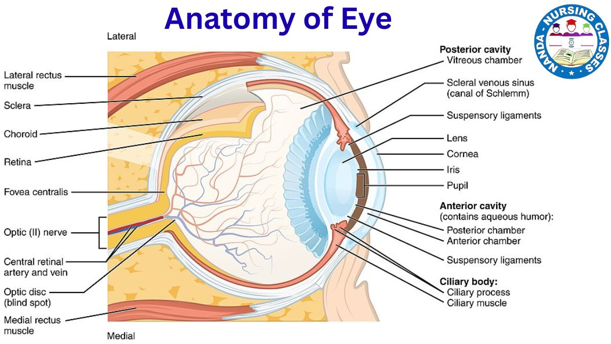
The eye is a spherical-shaped organ located within the orbit (eye socket). It is protected and supported by accessory structures such as the eyelids, eyelashes, and lacrimal glands. The average diameter of the eyeball is approximately 2.5 cm.
Anatomically, the eyeball is divided into two main parts:
-
The anterior part, which constitutes about 1/6th of the eyeball
-
The posterior part, which makes up the remaining 5/6th
The eye is made up of three main layers and several internal components that play a vital role in vision. These components work together to receive visual information, which is then transmitted to the brain through the optic nerve.
Layers of the Eye
The wall of the eyeball is made up of three main layers of tissue- the fibrous layer, the Vascular Layer, and the Retinal Layer, which are also referred to as the layers of the eye. These three layers are described below:
Fibrous Layer tissue (Outer Layer)
The fibrous layer, also known as the outer layer of the eye, is the outermost protective covering of the eyeball. It is composed of two main parts: the cornea and the sclera.
- Cornea: The cornea is the transparent, dome-shaped front part of the eye. It allows light to enter the eyeball and plays a vital role in bending (refracting) light rays to help focus them on the retina, contributing significantly to clear vision.
- Sclera: The sclera, often referred to as the “white of the eye,” is the opaque, tough outer covering that surrounds the rest of the eyeball. It provides structural support and protection, helping the eye maintain its shape and shielding the delicate inner components from injury.

Vascular Layer (Middle Layer or Uvea)
The vascular layer, also known as the middle layer or uvea, lies beneath the fibrous layer and is rich in blood vessels and pigment. It plays a crucial role in nourishing the eye and regulating its internal functions. This layer consists of the choroid, ciliary body, and iris.
- Choroid: The choroid is a darkly pigmented layer that lines most of the inner surface of the sclera. It contains numerous blood vessels that supply oxygen and nutrients to the retina, helping to maintain the health of the eye tissues.
- Ciliary Body: The ciliary body is located just behind the iris and contains muscle fibers that help adjust the shape of the lens. This adjustment is essential for focusing on objects at different distances, a process known as accommodation.
- Iris: The iris is the colored part of the eye and is responsible for controlling the size of the pupil. By adjusting the pupil size, the iris regulates the amount of light that enters the eye, protecting it from excessive brightness and improving focus.
Retinal Layer (Inner Layer)
The retinal layer, also known as the inner layer, is the innermost and most delicate part of the eye. It contains the retina, a light-sensitive layer made up of photoreceptor cells called rods and cones. These cells detect light and convert it into electrical signals, which are then transmitted to the brain through the optic nerve, allowing us to perceive images.
Internal Structures of the Eye
The internal structures of the eye work together to ensure proper vision by guiding and processing light. These structures include the lens, aqueous humor, vitreous humor, pupil, and optic nerve.
lens
The lens is a transparent and flexible structure located behind the iris. It plays a vital role in focusing light onto the retina by changing its shape depending on the distance of the object being viewed—a process known as accommodation.
Aqueous humor
The aqueous humor is a clear, watery fluid found in the anterior chamber of the eye, between the cornea and the lens. It helps to maintain intraocular pressure, nourishes the cornea and lens, and removes waste products.
Vitreous humor
The vitreous humor is a gel-like substance that fills the posterior chamber of the eye, located between the lens and the retina. It helps the eye maintain its spherical shape and provides internal support to keep the retina in place.
Pupil
The pupil is the central opening of the iris. It functions as a gateway for light, expanding or contracting to regulate the amount of light that enters the eye, depending on environmental brightness.
Optic nerve
The optic nerve is responsible for transmitting electrical signals from the retina to the brain, where they are interpreted as visual images. It serves as the communication link between the eye and the visual centers of the brain.
Physiology of the Eye
What is the physiology of the Eye ?
The physiology of the eye refers to the complex processes that allow us to see and interpret the world around us. While the structure of the eye provides the physical framework, its function depends on a series of finely coordinated actions that involve light transmission, photoreception, and image interpretation by the brain. Let’s learn one by one:
Light Transmission and Refraction
The process of vision begins when light enters the eye. The first structure it encounters is the cornea, a transparent, curved surface that bends (refracts) the incoming light rays. This initial refraction helps direct the light toward the lens.
The lens then performs fine adjustments by changing its shape, a process known as accommodation. This ensures that the light is precisely focused onto the retina, the light-sensitive layer at the back of the eye.
Meanwhile, the pupil, located at the center of the iris, acts as a light regulator. It dilates (enlarges) in dim light to allow more light in and constricts (shrinks) in bright light to reduce light entry. These changes in pupil size are controlled by the muscles of the iris, ensuring that the right amount of light reaches the retina for clear vision.
Let us understand in short Light Transmission and Refraction :
- Light enters through the cornea, which bends (refracts) it toward the lens.
- The lens further adjusts its shape (accommodation) to focus light onto the retina.
- The pupil dilates or constricts to regulate light entry, controlled by the iris muscles.
Photoreception and Signal Processing
Once light is focused onto the retina, the process of photoreception begins. The retina contains two types of specialized photoreceptor cells—rods and cones—each with distinct functions.
Rods are responsible for vision in low-light conditions and help detect shades of black, white, and gray. They are highly sensitive to light but do not detect color, making them essential for night vision.
Cones, on the other hand, function best in bright light and are responsible for color vision. They allow us to perceive fine details and a wide range of colors.
These photoreceptors convert light into electrical signals through a biochemical process. These signals are then processed by other retinal neurons and ultimately transmitted through the optic nerve to the brain, where they are interpreted as visual images.
Let us understand in short, Photoreception and Signal Processing :
- The retina contains two types of photoreceptor cells:
- Rods: Function in low-light conditions and provide black-and-white vision.
- Cones: Function in bright light and allow color vision.
- The photoreceptors convert light into electrical signals, which are transmitted via the optic nerve to the brain.
Image Formation and Interpretation
After the retina converts light into electrical signals, these signals are transmitted through the optic nerve to the visual cortex located in the occipital lobe of the brain. The visual cortex is responsible for processing and interpreting the incoming signals.
Although the image formed on the retina is inverted (upside down and reversed), the brain automatically corrects it, allowing us to perceive a clear, upright image of the world around us. This complex process enables us to recognize shapes, colors, depth, and motion, forming the basis of visual perception.
Let us understand in short, Image Formation and Interpretation
- The optic nerve carries signals to the visual cortex in the brain.
- The brain processes these signals to form a clear, upright image.
That’s all for now about the eye anatomy and physiology. If you have any questions or doubts, feel free to ask in the comment section below, or you can reach out to us directly through our social media accounts. We’re here to help you learn better!
We have divided the sensory organs into five sections for your convenience. You may read each part by clicking on the topics listed below:
Part 1 – Skin 🖐️
Part 2 – Eyes 👁️
Part 3 – Nose 👃
Part 4 – Ears👂
Part 5 – Tongue 👅
Common disorders of the eye
1. Myopia (Nearsightedness)
A refractive error where distant objects appear blurry because light focuses in front of the retina instead of on it. Often caused by an elongated eyeball or curved cornea.
2. Hyperopia (Farsightedness)
A condition in which nearby objects appear blurry while distant objects are seen clearly. It happens when light focuses behind the retina, typically due to a shorter eyeball.
3. Astigmatism
A common vision problem caused by an irregularly shaped cornea or lens, leading to blurred or distorted vision at all distances.
4. Presbyopia
An age-related condition in which the eye gradually loses the ability to focus on close objects due to hardening of the lens.
5. Cataract
Clouding of the eye’s natural lens, leading to blurry vision, glare, and faded colors. Most commonly occurs with aging but can also result from trauma or medication.
6. Glaucoma
A group of eye conditions that damage the optic nerve, usually due to increased intraocular pressure. If left untreated, it can lead to permanent vision loss.
7. Conjunctivitis (Pink Eye)
Inflammation or infection of the conjunctiva—the clear membrane covering the white part of the eye. It causes redness, irritation, and discharge, and can be bacterial, viral, or allergic.
8. Dry Eye Syndrome
Occurs when the eyes do not produce enough tears or the tears evaporate too quickly, leading to irritation, burning, or a gritty sensation.
9. Macular Degeneration
A progressive eye disease affecting the macula—the central part of the retina—resulting in central vision loss. Common in older adults.
10. Diabetic Retinopathy
A complication of diabetes that damages the small blood vessels in the retina. It can cause blurred vision, floaters, and even blindness if untreated.
11. Retinal Detachment
A medical emergency where the retina pulls away from its normal position. It may cause flashes of light, floaters, or a shadow over part of the visual field.
12. Amblyopia (Lazy Eye)
A developmental problem where one eye fails to achieve normal visual acuity. It usually begins in childhood and must be treated early.
13. Strabismus (Crossed Eyes)
A condition where both eyes do not align properly and may point in different directions. It can affect depth perception and may lead to amblyopia if untreated.
14. Color Blindness
A genetic disorder where a person has difficulty distinguishing certain colors, most commonly red and green.
Key Terms Glossary 🔑 : Eye Anatomy and Physiology
Eye Anatomy
The study of the physical structures of the eye.
Eye Physiology
The study of how the eye functions to produce vision.
Cornea
The transparent front surface of the eye that bends light for focusing.
Sclera
The white outer layer of the eye that gives shape and protection.
Iris
The colored part of the eye that controls the size of the pupil.
Pupil
The opening in the center of the iris that regulates light entry.
Lens
A clear, flexible structure that focuses light onto the retina.
Retina
The inner layer of the eye containing photoreceptors (rods and cones).
Rods
Photoreceptor cells that detect light and function in dim lighting.
Cones
Photoreceptor cells responsible for color vision and detail in bright light.
Optic Nerve
The nerve that carries visual information from the retina to the brain.
Aqueous Humor
Clear fluid in the front chamber of the eye; maintains pressure and nourishes.
Vitreous Humor
Jelly-like fluid filling the back of the eye; helps maintain eye shape.
Choroid
A layer filled with blood vessels between the retina and sclera.
Ciliary Body
A structure containing muscles that change the lens shape for focusing.
Accommodation
The process by which the lens changes shape to focus on near/far objects.
Photoreceptors
Cells (rods and cones) in the retina that convert light into electrical signals.
Visual Cortex
The part of the brain that processes visual information.
Myopia
Nearsightedness; difficulty seeing distant objects clearly.
Hyperopia
Farsightedness; difficulty seeing nearby objects clearly.
Lacrimal Glands
Glands that produce tears to keep the eye moist and clean.
Orbit
The bony socket in the skull that houses and protects the eyeball.
if you want to know anything just comment it
You can save the image of terminology or maybe download a PDF

Eye anatomy and physiology pdf
If you want Eye Anatomy and Physiology pdf and Eye Anatomy and Physiology ppt, click the button given below and downlod it.
FAQs on Anatomy and Physiology of the Eye
1. What are the main parts of the eye?
The eye has several major structures:
-
Cornea – the clear, outer surface that lets light in.
-
Iris – the colored part that controls the size of the pupil.
-
Pupil – the black opening that adjusts to let more or less light in.
-
Lens – focuses light onto the retina.
-
Retina – contains photoreceptor cells (rods and cones) that detect light and color.
-
Optic Nerve – carries visual information to the brain.
2. How does the eye work to produce vision?
Light enters through the cornea and pupil, passes through the lens, and is focused onto the retina. The retina’s photoreceptors convert the light into electrical signals, which travel via the optic nerve to the brain, where the image is interpreted.
3. What are rods and cones?
-
Rods detect black and white and are useful in low-light conditions.
-
Cones detect color and function best in bright light.
4. What is the function of the cornea?
The cornea protects the eye and helps bend (refract) light to focus it on the retina.
5. How does the lens adjust focus?
The lens changes shape through a process called accommodation, becoming thicker for near objects and thinner for distant ones.
6. What is the aqueous humor?
It’s a clear fluid in the front part of the eye that helps maintain pressure, provides nutrients, and removes waste.
7. What is the vitreous humor?
A jelly-like substance that fills the space between the lens and the retina, helping the eye maintain its shape.
8. What causes common vision problems like myopia and hyperopia?
-
Myopia (nearsightedness) – occurs when the eyeball is too long, causing images to focus in front of the retina.
-
Hyperopia (farsightedness) – happens when the eyeball is too short, causing images to focus behind the retina.
9. What is the role of the optic nerve?
The optic nerve transmits visual information from the retina to the brain.
10. Why is understanding the eye important in healthcare?
Nurses and healthcare workers need to assess vision, detect signs of disease (like glaucoma or cataracts), and provide care that considers a patient’s ability to see.
If you liked this post, please share it with others. Feel free to leave a comment below if you have any questions or feedback!


I¦ve been exploring for a little bit for any high-quality articles or weblog posts on this kind of area . Exploring in Yahoo I at last stumbled upon this website. Reading this information So i¦m glad to express that I have a very good uncanny feeling I came upon exactly what I needed. I such a lot certainly will make sure to do not fail to remember this web site and provides it a glance on a continuing basis.
This is the right blog for anyone who wants to find out about this topic. You realize so much its almost hard to argue with you (not that I actually would want…HaHa). You definitely put a new spin on a topic thats been written about for years. Great stuff, just great!