The ear is one of the five primary sensory organs and plays a vital role in two major functions: hearing (auditory perception) and balance (equilibrium). Understanding the Anatomy and Physiology of the Ear is crucial for nursing students, medical aspirants, and healthcare professionals, as it provides a clear foundation in how sound is detected, processed, and how body balance is maintained.
🦻 Anatomy and Physiology of the Ear {Hearing and Balance System}
What is the anatomy and physiology of ear?
In the study of human anatomy and physiology, the ear is classified as part of the special senses and is anatomically divided into three main sections: the outer ear, middle ear, and inner ear—each with specialized structures that work in coordination to transmit sound waves and regulate equilibrium.
Anatomy of the Ear
What is the anatomy of ear?
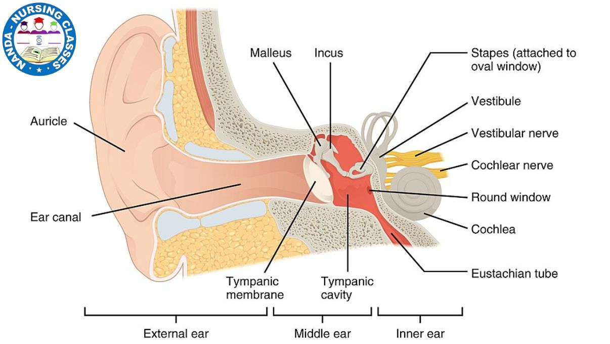
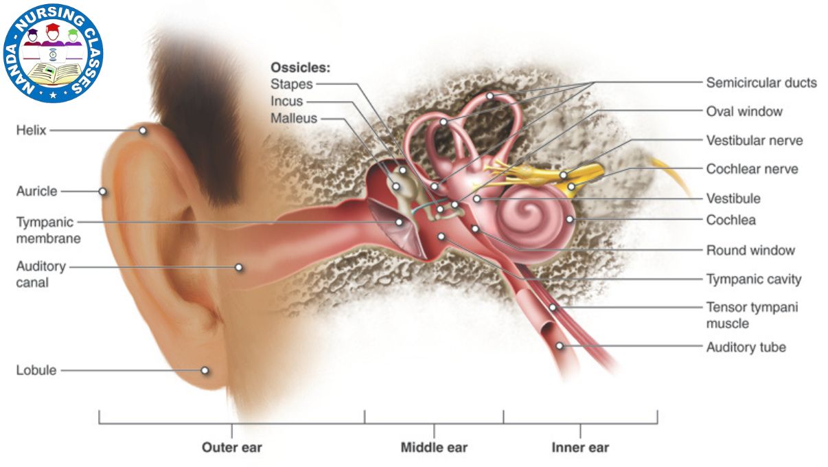
The ear is a complex sensory organ divided into three anatomical regions—each performing essential roles in hearing and balance. These are the external (outer) ear, middle ear, and internal (inner) ear. Understanding the anatomy of the ear helps explain how sound waves travel and are converted into electrical signals the brain can interpret.
External Ear (Outer Ear)
The external ear is the first part of the auditory system that interacts with the environment. It is composed of the pinna (auricle) and the external auditory canal.
-
The pinna, which is the visible part of the ear, acts like a funnel. It collects sound waves from the environment and channels them into the auditory canal.
-
These sound waves travel through the auditory canal and hit the tympanic membrane (commonly known as the eardrum).
-
The tympanic membrane serves as a boundary between the external and middle ear. It vibrates in response to the incoming sound waves, beginning the process of mechanical sound transmission.
Middle Ear
The middle ear is an air-filled cavity that contains three tiny bones known as ossicles. These are the malleus (hammer), incus (anvil), and stapes (stirrup).
-
These ossicles work together to amplify and transfer the vibrations from the tympanic membrane to the oval window, which is the entrance to the inner ear.
-
The malleus is attached to the eardrum and passes vibrations to the incus, which then transmits them to the stapes.
-
The stapes, being the smallest bone in the human body, transmits the vibration to the fluid-filled cochlea in the inner ear.
-
The Eustachian tube, also located in the middle ear, connects it to the throat (nasopharynx). This tube helps equalize air pressure on both sides of the eardrum, ensuring proper vibration and preventing damage or discomfort (like during altitude changes).
Inner Ear
The inner ear, also called the labyrinth, is a complex structure responsible for both hearing and balance. It contains three major components:
-
Cochlea – This spiral-shaped organ is filled with fluid and contains the organ of Corti, the actual hearing organ. Inside the organ of Corti are hair cells that move in response to fluid vibrations. These movements are converted into nerve impulses, which travel to the brain via the vestibulocochlear nerve (Cranial Nerve VIII).
-
Vestibule – Located in the central part of the inner ear, the vestibule helps detect linear changes in head position, contributing to our sense of balance.
-
Semicircular Canals – These three looped structures are oriented in different planes and help detect rotational movements of the head. They send signals to the brain that help maintain balance during motion.
Together, these structures ensure that we can hear clearly and maintain our equilibrium while standing, walking, or moving.
Physiology of the Ear
What is the physiology of ear?
The ear is not just an organ for hearing—it also plays a critical role in maintaining balance. The physiology of the ear involves a series of processes that convert sound waves into electrical signals and send them to the brain, while also detecting changes in body position and motion. These functions are made possible by the auditory system and the vestibular system, both located in the inner ear. Hearing allows us to perceive and interpret sound waves from the environment, while balance ensures we can stand, walk, and move without falling. In this section, we explore how sound waves are transformed into nerve impulses and how the inner ear helps us maintain balance, even with sudden movements.
🔊 A. Hearing Process
The hearing process is a complex yet fascinating sequence that allows us to perceive sounds from our surroundings. It involves the transformation of sound waves into electrical signals that the brain can interpret.
-
Sound wave entry:
Sound waves from the environment enter through the external ear and travel down the auditory canal. -
Tympanic membrane vibration:
These waves strike the tympanic membrane (eardrum), causing it to vibrate. -
Ossicles amplify the sound:
Vibrations are transferred to the middle ear bones (ossicles):-
Malleus (hammer)
-
Incus (anvil)
-
Stapes (stirrup)
These bones act like a lever system to amplify the sound vibrations.
-
-
Transmission to the inner ear:
The stapes transmits these amplified vibrations to the oval window, a membrane leading into the cochlea of the inner ear. -
Fluid movement in the cochlea:
Vibrations enter the fluid-filled cochlea, creating waves in the endolymph. -
Stimulation of hair cells:
The movement of fluid stimulates hair cells in the organ of Corti (inside the cochlea). These hair cells are specialized sensory receptors. -
Signal transmission to the brain:
Hair cells convert the mechanical vibrations into electrical nerve impulses, which travel via the auditory nerve (Cranial Nerve VIII) to the brain’s auditory cortex, where they are processed and recognized as sound.
⚖️ B. Balance and Equilibrium
The vestibular system, located in the inner ear, is responsible for maintaining balance and spatial orientation. It consists of the semicircular canals and vestibule (utricle and saccule).
-
Detection of head movement:
The semicircular canals are filled with a fluid called endolymph. When the head rotates, the fluid shifts, causing movement of hair cells in the canal linings. This detects rotational motion. -
Detection of linear movement and gravity:
The utricle and saccule (part of the vestibule) detect changes in linear movement (e.g., walking forward) and gravity (e.g., tilting the head). Tiny crystals (otoliths) inside these structures move in response to head position, stimulating sensory hair cells. -
Signal transmission for coordination:
The information from these receptors travels via the vestibular nerve (a branch of the vestibulocochlear nerve, CN VIII) to the brainstem and cerebellum, where it is processed to help maintain posture, balance, and coordinated movement.
Vestibular organ
What is the vestibular organ ?
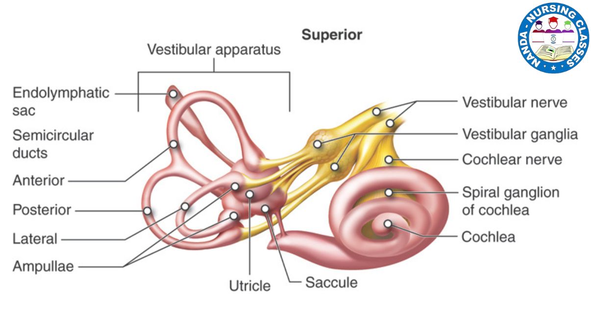
The vestibular organ is a specialized structure located in the inner ear. Its primary role is to help maintain balance, detect head movement, and provide the brain with information about spatial orientation. In simple terms, it is the organ of balance in the ear, working silently to keep our body upright and steady during movement.
1. 📍 Location and Structure
-
The vestibular organ is found deep within the inner ear, enclosed in a bony structure called the bony labyrinth, which is part of the temporal bone of the skull.
-
It is closely associated with the cochlea, which is responsible for hearing.
-
The vestibular system itself includes two main parts:
-
Semicircular canals – three looped tubes that detect rotational movements.
-
Otolith organs – the utricle and saccule, which detect linear motion and head position related to gravity.
-
2. 🔄 Semicircular Canals
-
There are three semicircular canals, each positioned at roughly right angles to each other, allowing detection of movement in three-dimensional space:
-
Anterior (superior) canal – Detects nodding movements of the head (up and down).
-
Posterior canal – Detects tilting movements from side to side.
-
Lateral (horizontal) canal – Detects rotation of the head (turning side to side, like shaking your head “no”).
-
-
Inside each canal is a fluid called endolymph. As the head moves, the fluid shifts, and a sensory structure called the crista ampullaris inside each ampulla (enlarged base of each canal) detects this motion.
3. ↕️ Otolith Organs: Utricle and Saccule
-
The otolith organs are responsible for detecting linear acceleration and head position in relation to gravity.
-
Each organ contains:
-
A patch of hair cells embedded in a gelatinous layer.
-
On top of this gel layer are small crystals called otoliths (made of calcium carbonate).
-
-
When the head moves, the otoliths shift due to gravity, bending the hair cells and sending signals to the brain.
-
The utricle detects horizontal movements (such as walking forward or backward).
-
The saccule detects vertical movements (such as jumping or nodding).
-
4. 🧠 Nerve Supply
-
The vestibulocochlear nerve (Cranial Nerve VIII) carries signals from the vestibular organs to the brainstem.
-
These signals are processed by the brain to maintain:
-
Balance
-
Eye coordination
-
Postural adjustments
-
-
The integration of this data helps the body react appropriately to changes in movement and head position, preventing dizziness and falls.
⚙️ Physiology of the Vestibular Organ (Vestibular Function)
How does the vestibular system work ?
The vestibular organ plays a crucial role in helping the body maintain balance, spatial orientation, and coordination. Located in the inner ear, it works in harmony with the visual system and proprioceptive system (sensors in muscles and joints) to provide information about head position and motion. Below are the key physiological functions of the vestibular system:
1. 🧍♂️ Balance and Spatial Orientation
-
The vestibular system constantly monitors the position and movement of the head in relation to gravity.
-
It communicates with the visual system (eyes) and proprioceptors (receptors in muscles and joints) to help you maintain equilibrium, especially during movement.
-
For example, when you tilt your head or move suddenly, the vestibular system detects this change and sends signals to your brain to adjust posture and keep you upright.
2. 🔁 Reflexes and Coordination
The vestibular system is responsible for several reflexes that help maintain stability:
a. Vestibulo-Ocular Reflex (VOR)
-
This reflex ensures that your eyes remain fixed on an object even when your head is moving.
-
For example, when you’re walking or turning your head, the VOR keeps your vision stable by adjusting your eye movements automatically.
b. Vestibulospinal Reflex
-
This reflex helps you maintain posture and body balance.
-
It sends signals to muscles in your body to adjust your stance and prevent falls when your head position changes unexpectedly.
3. 🌀 Motion Detection
The vestibular system contains specialized structures that detect different types of motion:
a. Semicircular Canals – Detect Rotational Movement
-
The three semicircular canals sense rotational motion of the head (such as spinning, turning, or tilting).
-
When you rotate your head, fluid (endolymph) inside the canals shifts and stimulates sensory hair cells in the crista ampullaris, triggering nerve signals.
b. Otolith Organs – Detect Linear Acceleration and Tilting
-
The utricle and saccule detect changes in straight-line movement and head tilt.
-
Small crystals (otoliths) inside these organs shift with gravity or motion, bending the hair cells beneath them and sending signals to the brain.
Together, the structures of the vestibular organ provide vital feedback that enables us to:
-
Walk steadily
-
Stand upright
-
Coordinate head and eye movements
-
Adjust to moving environments, like riding in a car or standing on a moving train
✅ Conclusion
The ear is a vital sensory organ that plays a dual role in hearing and maintaining balance. Through its three anatomical regions—the outer ear, middle ear, and inner ear—the ear captures sound waves, amplifies them, and converts them into electrical impulses that are interpreted by the brain. Simultaneously, the vestibular system within the inner ear continuously monitors head position and motion, working in coordination with the eyes and muscles to maintain equilibrium and spatial orientation.
Understanding the anatomy and physiology of the ear provides crucial insights into how we perceive our environment and maintain body posture. For nursing students and healthcare professionals, this knowledge lays the foundation for diagnosing and managing conditions related to hearing loss, vertigo, and balance disorders. Mastery of this topic enhances both academic learning and clinical competency.
🔍 Frequently Asked Questions (FAQs)
❓1. What are the main parts of the human ear?
The human ear is divided into three main parts:
-
Outer ear (pinna and auditory canal)
-
Middle ear (ossicles: malleus, incus, stapes)
-
Inner ear (cochlea and vestibular system)
Each part plays a unique role in the hearing and balance processes.
❓2. How does the ear help in maintaining balance?
The vestibular organ, located in the inner ear, detects head position and motion. It sends signals to the brain to maintain balance, spatial orientation, and postural control using the semicircular canals, utricle, and saccule.
❓3. What is the function of the cochlea in the ear?
The cochlea is a spiral-shaped organ in the inner ear responsible for hearing. It converts sound wave vibrations into electrical impulses, which are sent to the brain via the auditory nerve.
❓4. What is the role of the semicircular canals?
The semicircular canals are three looped structures that detect rotational movements of the head. They help the body maintain balance and eye coordination through the vestibulo-ocular reflex (VOR).
❓5. How do sound waves travel through the ear?
Sound waves follow this path:
Outer ear → Tympanic membrane → Ossicles → Oval window → Cochlea → Auditory nerve → Brain.
Each structure amplifies and processes sound for accurate hearing.
❓6. What is the vestibulocochlear nerve (Cranial Nerve VIII)?
The vestibulocochlear nerve carries signals from the cochlea (for hearing) and the vestibular system (for balance) to the brain. It plays a key role in both auditory perception and equilibrium control.
❓7. What happens if the vestibular system is damaged?
Damage to the vestibular system can lead to dizziness, vertigo, loss of balance, and nausea. This condition may result from infections, inner ear disorders, or head injuries.
❓8. Why is understanding ear anatomy important in nursing?
Nursing students must understand ear anatomy to recognize and manage conditions like ear infections, hearing loss, vertigo, and balance disorders, ensuring better patient care.
❓9. How do the otolith organs work?
The utricle and saccule contain crystals (otoliths) that shift in response to gravity and movement. These shifts bend hair cells and send signals to the brain about linear acceleration and head position.
❓10. Can hearing and balance be affected together?
Yes. Since both systems share the inner ear and vestibulocochlear nerve, issues like labyrinthitis or Meniere’s disease can affect both hearing and balance simultaneously.
That’s all for now about the Ear anatomy and physiology. If you have any questions or doubts, feel free to ask in the comment section below, or you can reach out to us directly through our social media accounts. We’re here to help you learn better!
We have divided the sensory organs into five sections for your convenience. You may read each part by clicking on the topics listed below:
Part 1 – Skin 🖐️
Part 2 – Eyes 👁️
Part 3 – Nose 👃
Part 4 – Ears👂
Part 5 – Tongue 👅
🔑 Key Terms Glossary: Ear Anatomy and Physiology
Pinna (Auricle)
The visible part of the outer ear that collects sound waves and funnels them into the auditory canal.
Auditory Canal
A tube-like structure that leads from the pinna to the eardrum; helps transmit sound waves inward.
Tympanic Membrane (Eardrum)
A thin membrane that vibrates when sound waves strike it, separating the outer ear from the middle ear.
Ossicles
Three small bones in the middle ear—malleus, incus, and stapes—responsible for amplifying sound vibrations.
Malleus (Hammer)
The first ossicle bone, attached to the tympanic membrane, that receives sound vibrations and transmits them to the incus.
Incus (Anvil)
The second ossicle bone that connects the malleus to the stapes and helps transmit vibrations.
Stapes (Stirrup)
The smallest bone in the human body; passes vibrations from the incus to the oval window of the cochlea.
Eustachian Tube
A narrow canal that connects the middle ear to the throat (nasopharynx); equalizes air pressure across the eardrum.
Cochlea
A spiral-shaped, fluid-filled organ in the inner ear that transforms sound vibrations into electrical signals.
Organ of Corti
Located within the cochlea, this structure contains hair cells that act as sensory receptors for hearing.
Hair Cells
Specialized cells in the cochlea and vestibular system that convert mechanical movements into electrical nerve impulses.
Endolymph
A fluid inside the cochlea and semicircular canals that helps carry vibrations and motion signals to sensory cells.
Vestibular System
Part of the inner ear responsible for balance and spatial orientation, composed of semicircular canals and otolith organs.
Semicircular Canals
Three looped tubes (anterior, posterior, and lateral) that detect head rotation in different directions.
Crista Ampullaris
A sensory receptor inside each semicircular canal’s ampulla that detects changes in head rotation.
Otolith Organs
Includes the utricle and saccule, which detect linear movement and head position relative to gravity.
Utricle
A component of the vestibule that senses horizontal motion, like moving forward or backward.
Saccule
A vestibular organ that detects vertical motion, such as jumping or moving up/down.
Otoliths
Tiny calcium carbonate crystals in the otolith organs that respond to gravity and movement, stimulating hair cells.
Vestibulocochlear Nerve (Cranial Nerve VIII)
The eighth cranial nerve that transmits auditory signals from the cochlea and balance information from the vestibular system to the brain.
Vestibulo-Ocular Reflex (VOR)
A reflex that stabilizes vision by adjusting eye movements to compensate for head movements.
Vestibulospinal Reflex
A reflex that helps maintain balance and posture by activating muscles in response to head motion.
Auditory Cortex
Part of the brain’s temporal lobe where sound signals are processed and interpreted as hearing.
if you want to know anything just comment it
You can save the image of terminology or maybe download a PDF

If you liked this post, please share it with others. Feel free to leave a comment below if you have any questions or feedback!