The cardiovascular system also known as the circulatory system, is essential for maintaining the body’s homeostasis. It is responsible for transporting oxygen, nutrients, hormones, and waste products throughout the body. In the cardiovascular system anatomy and physiology we mainly learn about four things about the circulatory system is Heart, Blood, Vessels, and lymph. Below are the key components and concepts of the circulatory system explained for nursing students. When you are studying or learning about the Cardiovascular System it is called
💡 “Angiology” is the study of the cardiovascular system, and William Harvey is called the “Father of Angiology” for discovering it in 1628.
Cardiovascular system or circulatory system
❤️ What is the Cardiovascular System (Circulatory System)?
The cardiovascular system, also known as the circulatory system, is a vital organ system in the human body responsible for transporting blood, oxygen, nutrients, hormones, and waste products to and from the body’s tissues.
Types of the Cardiovascular System
There are two types of Cardiovascular System which is the open Cardiovascular System and the closed Cardiovascular System. Open Cardiovascular Systems means circulation in any ducts or veins of mammals and Closed Cardiovascular System means circulation without ducts or veins. Examples include Butterflies, insects, octopuses, & cockroaches. But in nursing, we only learn about the Human Cardiovascular System only, Let’s learn in detail about the Cardiovascular System.
In the study of the anatomy and physiology of the cardiovascular system, we primarily focus on four main components: the heart, blood, blood vessels, and the lymphatic system.
Heart
The heart is a hollow muscular organ of the body, The heart’s main function is to pump blood all over the body, The Heart pumps around 7200 liters of blood in a day. Heart beats in a day about 100000 times. Heart weight in males is approximately 310 grams and in females, it has approximately. 255 grams. The heart lies obliquely, its base above and apex below. It is basically all about the Heart let’s learn in detail the Structure & Position of the Heart, Functions of the Heart, the Conduction System, the Cardiac Cycle, normal values of the heart, and more.
Structure of the Heart
Layers of Heart
The heart is a muscular, cone-shaped organ the pericardium encloses it, a double-layered sac that protects and anchors it. If we talk about heart layer it has three layers endocardium, myocardium, and pericardium. The endocardium is the Inner layer, the Myocardium is the Muscular middle layer, responsible for contraction and the Epicardium is the Outer layer, part of the pericardium.
Chambers of Heart
As you learned the heart is a hollow organ but it’s not completely hollow it is divided into four chambers, we know them by their position right and left, and their functional type. The heart has two types of chambers, each with distinct functions and names: atrium and ventricle. Here’s why they are called so:
Atrium:
- The word “atrium” originates from the Latin word ātrium, meaning “entrance hall” or “central room.”
- In the heart, the atrium serves as the “entrance hall” for blood, receiving it from the body or lungs and holding it temporarily before it moves to the ventricles.
- Humans have two atria:
-
- The right atrium receives deoxygenated blood from the body.
- The left atrium receives oxygenated blood from the lungs.
-
Ventricle:
- The term “ventricle” comes from the Latin word ventriculus, meaning “small belly” or “chamber.”
- Ventricles are responsible for pumping blood out of the heart, either to the lungs (right ventricle) or the rest of the body (left ventricle). These chambers have thicker walls compared to the atria because they need to generate greater force to propel the blood.
In short, the atria are entry chambers for blood, while the ventricles are the strong pumping chambers, and their names reflect their functions and anatomical structure.
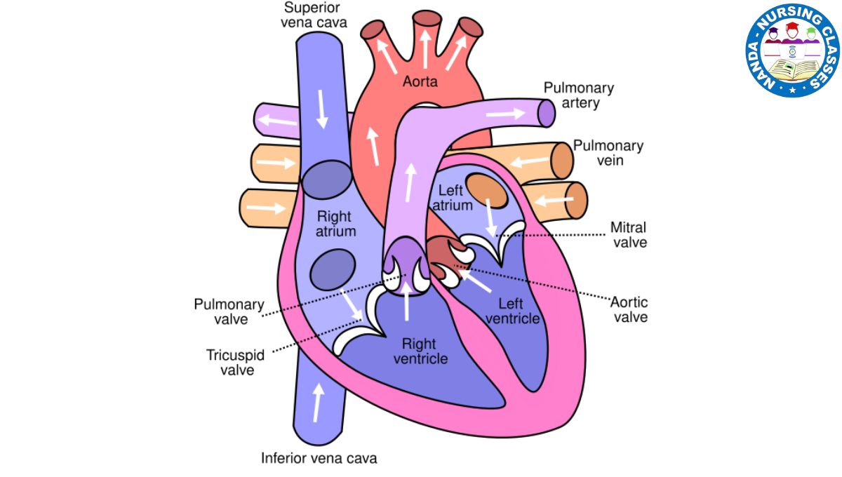
The heart has four chambers:
- Right atrium (RA): Receives deoxygenated blood from the body.
- Right ventricle (RV): Pumps deoxygenated blood to the lungs via the pulmonary artery.
- Left atrium (LA): Receives oxygenated blood from the lungs.
- Left ventricle (LV): Pumps oxygenated blood to the body through the aorta.
Valves of the heart
The heart has also four valves, the valves mean they have the capability to allow and stop the flow of a pathway, in the heart the valves prevent the backflow of blood, and they allow it to flow in the same direction which we will learn in Circulation of Blood.
- Atrioventricular valves: Tricuspid (right) and mitral (left).
- Semilunar valves: Pulmonary (right) and aortic (left).
Position of the Heart
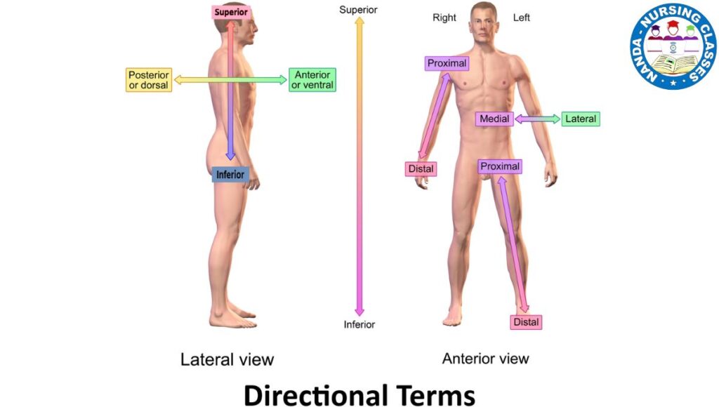
The heart is located in the mediastinum between the lungs on top of the diaphragm, behind the sternum, in front of thoracic vertebrae, and protected by ribs.1/3 position of the heart is in the right side of the midline of the body and 2/3 is left side of the midline of the body. As you know normally Heart is located on the left side of the body is called levocardia but if the Heart is located on the right side of the body is known as Dextrocardia due to a birth defect.
Functions of the Heart
Functions of the Heart
The heart is a vital organ responsible for maintaining blood circulation throughout the body. Its primary functions include:
- Pumping Blood
- Circulating Oxygen and Nutrients
- Removing Waste Products
- Maintaining Blood Pressure
- Regulating flow
- Facilitating Hormone Delivery
- Ensuring Separation of Oxygenated and Deoxygenated Blood
- Supporting Thermoregulation
1. Pumping Blood
The heart pumps oxygenated blood from the lungs to the rest of the body through the left ventricle. It pumps deoxygenated blood from the body to the lungs for oxygenation through the right ventricle.
2. Circulating Oxygen and Nutrients:
Blood carries oxygen and essential nutrients to tissues and organs, ensuring they function properly.
3. Removing Waste Products:
The heart helps transport waste products, such as carbon dioxide and metabolic byproducts, to organs like the lungs, kidneys, and liver for elimination.
4. Maintaining Blood Pressure:
By contracting and relaxing in a rhythmic manner, the heart generates and regulates blood pressure, ensuring adequate blood flow to all parts of the body.
5. Regulating flow
Ensures unidirectional blood flow using valves.
6. Facilitating Hormone Delivery:
The heart helps circulate hormones produced by glands, which regulate various bodily processes like metabolism, growth, and stress response.
7. Ensuring Separation of Oxygenated and Deoxygenated Blood:
The heart’s four-chambered structure ensures that oxygen-rich blood does not mix with oxygen-poor blood, maintaining efficient circulation.
8. Supporting Thermoregulation:
By controlling blood flow to the skin, the heart helps regulate body temperature, dissipating heat when necessary.
These functions collectively sustain life by delivering essential resources to the body and removing harmful substances.
Conduction System of the Heart
The conduction system initiates and regulates the heartbeat, ensuring the heart functions as an efficient pump. The conduction system of the heart is a network of specialized cells responsible for generating and transmitting electrical impulses. These impulses ensure the heart beats in a coordinated and rhythmic manner to pump blood effectively. Here’s a simplified explanation:
- Sinoatrial (SA) Node:
- Known as the natural “pacemaker” of the heart, the SA node is located in the right atrium.
- It initiates electrical signals that cause the atria to contract, pushing blood into the ventricles.
- These impulses set the heart rate.
- Atrioventricular (AV) Node:
- Situated between the atria and ventricles, the AV node acts as a relay station.
- It delays the electrical signal slightly, allowing the ventricles enough time to fill with blood before contracting.
- Bundle of His:
- After the AV node, the impulse travels to the Bundle of His, a pathway located in the septum (wall) between the ventricles.
- This bundle splits into right and left branches, directing the signal to both ventricles.
- Purkinje Fibers:
- These fibers spread the electrical impulse throughout the ventricular walls.
- This causes the ventricles to contract, pumping blood to the lungs (right ventricle) and the rest of the body (left ventricle).
This entire process happens rapidly and repeatedly, ensuring the heart maintains a steady rhythm and circulates blood effectively. The coordination of the conduction system is critical for maintaining life.
Cardiac Cycle
The cardiac cycle involves one complete heartbeat, The cardiac cycle consists of two phases Systole and Diastole, which means contraction and relaxation or we can say Depolarization and Repolarization,
Is this a complicated terminology? Let’s simplify it by understanding the meaning of each term.
Systole = contraction = Depolarization – All three have the same meaning, which means the blood is ejected from the heart, and
Diastole = relaxation = Repolarization – All three have the same meaning, which means the heart chambers refill with blood.
The cardiac cycle is completed in 0.8 seconds, as we know in one cardiac cycle there is a two-phase contraction and relaxation of heart muscles so in 0.8 seconds contraction takes its own time and relaxation takes its own time are explained below:
- Atrial Depolarization – 0.1 Second
- Ventricular Depolarization – 0.3 Second
- Atrial Repolarization – 0.5 Second
- Ventricular Repolarization – 0.7 Second
let’s learn in detail The cardiac cycle :
The cardiac cycle refers to the sequence of events that occur during one complete heartbeat. It includes the contraction and relaxation of the heart chambers to pump blood effectively. The cycle has two main phases: systole (contraction) and diastole (relaxation).
Stages of the Cardiac Cycle:
- Atrial Systole:
- The atria contract, pushing blood into the ventricles through the open atrioventricular (AV) valves (tricuspid on the right and mitral on the left).
- During this phase, the ventricles are relaxed and ready to receive blood.
- Ventricular Systole:
- After the atria relax, the ventricles contract. This contraction forces the AV valves to close, preventing backflow into the atria.
- As pressure builds in the ventricles, the semilunar valves (pulmonary valve on the right and aortic valve on the left) open, allowing blood to be ejected:
- The right ventricle pumps deoxygenated blood to the lungs via the pulmonary artery.
- The left ventricle pumps oxygenated blood to the body through the aorta.
- Diastole (Relaxation Phase):
- Both the atria and ventricles relax, allowing blood to fill the heart again.
- The semilunar valves close to prevent blood from flowing back into the ventricles.
- Blood flows passively from the veins (vena cava and pulmonary veins) into the atria and partially into the ventricles as the cycle starts again.
Key Points:
- The cardiac cycle is regulated by electrical signals generated in the heart’s conduction system, starting at the sinoatrial (SA) node, often called the pacemaker.
- The cycle ensures the heart efficiently pumps oxygen-rich blood to the body and oxygen-poor blood to the lungs for reoxygenation.
- A complete cardiac cycle takes about 0.8 seconds in a person with an average heart rate of 75 beats per minute.
The rhythmic and coordinated activity of the cardiac cycle ensures continuous blood circulation, sustaining life.
Blood
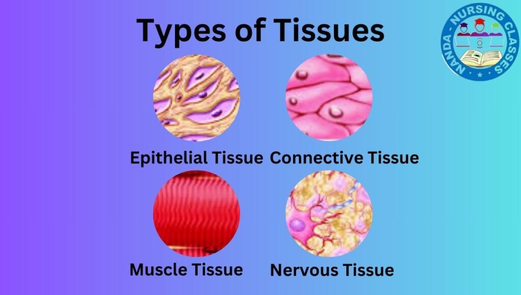
Blood is defined as a specialized connective tissue composed of plasma (the liquid component) and formed elements (RBCs, WBCs, and platelets) that circulate through the cardiovascular system to perform various functions, including transportation, regulation, and protection.
All about blood we learned in the previous post if you want full details of blood click here to learn more.
Blood Vessels
Blood vessels are an essential part of the cardiovascular system. They form a complex network of tubes that transport blood throughout the body, ensuring that oxygen, nutrients, hormones, and waste products are delivered or removed efficiently.
There are three main types of blood vessels:
-
Arteries
-
Arteries carry oxygen-rich blood away from the heart to the body’s tissues (except for pulmonary arteries, which carry deoxygenated blood to the lungs).
-
They have thick, muscular walls to withstand high pressure from the heart’s pumping action.
-
-
Veins
-
Veins carry oxygen-poor blood back to the heart (except for pulmonary veins, which carry oxygenated blood from the lungs to the heart).
-
They have thinner walls and often contain valves to prevent backflow of blood, especially in the limbs.
-
-
Capillaries
-
Capillaries are the smallest blood vessels, connecting arteries and veins.
-
Their thin walls allow for exchange of gases, nutrients, and waste products between blood and surrounding tissues.
-
Together, these vessels form a closed-loop system that keeps blood circulating efficiently to support all the organs and tissues of the body.
Lymph
Lymph is a clear, colorless fluid that circulates through the lymphatic system, playing a vital role in immune defense and maintaining fluid balance in the body.
What is Lymph?
Lymph is formed when interstitial fluid (the fluid that surrounds body tissues) enters the lymphatic capillaries. It closely resembles plasma but contains fewer proteins.
Components of Lymph:
-
Water: Makes up most of the lymph fluid.
-
White blood cells (mainly lymphocytes): Key players in immune response.
-
Proteins and fats: Especially absorbed from the digestive system (called chyle).
-
Cellular waste and debris
Functions of Lymph:
-
Maintains Fluid Balance:
-
Returns excess tissue fluid to the bloodstream, preventing swelling (edema).
-
-
Supports Immune Function:
-
Transports white blood cells and filters harmful pathogens through lymph nodes.
-
-
Fat Absorption:
-
Absorbs fats and fat-soluble vitamins from the small intestine via specialized lymph vessels called lacteals.
-
Lymphatic Vessels and Nodes:
-
Lymphatic Vessels: Thin-walled vessels that carry lymph fluid toward the heart.
-
Lymph Nodes: Small, bean-shaped structures that filter lymph and trap bacteria, viruses, and other harmful substances.
-
Commonly found in the neck, armpits, chest, abdomen, and groin.
-
🩺 Diseases of the Cardiovascular System
The cardiovascular system is essential for delivering oxygen and nutrients to tissues. When any part of this system is damaged or does not function properly, it can lead to serious health conditions. Below are common cardiovascular diseases (CVDs) every nursing student should know:
Hypertension
A condition characterized by persistently high blood pressure in the arteries, increasing the risk of heart disease and stroke.
Coronary Artery Disease (CAD)
A disease caused by the narrowing or blockage of coronary arteries due to plaque buildup, reducing blood flow to the heart muscle.
Heart Failure
A condition where the heart is unable to pump blood effectively to meet the body’s needs.
Myocardial Infarction (Heart Attack)
Occurs when blood flow to a part of the heart is blocked, causing damage or death to heart muscle tissue.
Arrhythmia
An abnormal heart rhythm, which may be too fast, too slow, or irregular.
Valvular Heart Disease
Damage or dysfunction of one or more heart valves that affects the normal flow of blood through the heart.
Congenital Heart Defects
Structural abnormalities of the heart present from birth, affecting normal heart function.
Peripheral Artery Disease (PAD)
A condition involving the narrowing of arteries in the limbs, leading to reduced blood flow and symptoms like leg pain.
Stroke
A sudden interruption of blood supply to the brain, resulting in brain cell damage or death.
Atherosclerosis
The buildup of fatty deposits (plaque) inside the arteries, causing them to narrow and harden.
Key Terms Glossary: Cardiovascular System
| Term | Definition / Explanation |
|---|---|
| Cardiovascular System | Also called the circulatory system, responsible for transporting blood, oxygen, nutrients, hormones, and waste throughout the body. |
| Angiology | The study of the cardiovascular system. William Harvey is the “Father of Angiology.” |
| Heart | A hollow muscular organ that pumps blood throughout the body. |
| Pericardium | A double-layered sac that encloses and protects the heart. |
| Endocardium | The inner layer of the heart. |
| Myocardium | The muscular middle layer responsible for heart contraction. |
| Epicardium | The outer layer of the heart, part of the pericardium. |
| Atrium (Atria) | Upper heart chambers that receive blood; right atrium receives deoxygenated blood, left atrium receives oxygenated blood. |
| Ventricle | Lower heart chambers that pump blood out; right ventricle pumps to lungs, left ventricle pumps to the body. |
| Heart Valves | Structures that prevent backflow of blood and maintain unidirectional flow: atrioventricular (tricuspid, mitral) and semilunar (pulmonary, aortic) valves. |
| Mediastinum | The central compartment of the thoracic cavity where the heart is located. |
| Levocardia | Normal position of the heart on the left side of the midline. |
| Dextrocardia | A birth defect where the heart is located on the right side of the body. |
| Cardiac Cycle | The sequence of heart contraction (systole) and relaxation (diastole) during one heartbeat. |
| Systole | Contraction phase of the cardiac cycle where blood is ejected from the heart. |
| Diastole | Relaxation phase of the cardiac cycle where heart chambers refill with blood. |
| Sinoatrial (SA) Node | The natural pacemaker of the heart, initiating electrical impulses for heartbeat. |
| Atrioventricular (AV) Node | Relays and delays the electrical signal to allow ventricles to fill before contracting. |
| Bundle of His | Pathway transmitting impulses from AV node to ventricles. |
| Purkinje Fibers | Fibers that spread electrical impulses through ventricles causing contraction. |
| Blood | Specialized connective tissue composed of plasma, red blood cells, white blood cells, and platelets. |
| Arteries | Blood vessels carrying oxygen-rich blood away from the heart (except pulmonary arteries). |
| Veins | Blood vessels carrying oxygen-poor blood to the heart (except pulmonary veins). |
| Capillaries | Smallest blood vessels allowing exchange of gases and nutrients between blood and tissues. |
| Lymph | Clear fluid circulating in the lymphatic system, important for immune function and fluid balance. |
| Lymph Nodes | Small organs that filter lymph and trap pathogens. |
| Hypertension | Persistently high blood pressure increasing risk of heart disease and stroke. |
| Coronary Artery Disease (CAD) | Narrowing or blockage of coronary arteries reducing blood flow to heart muscle. |
| Heart Failure | Condition where the heart cannot pump enough blood to meet body’s needs. |
| Myocardial Infarction | Heart attack; damage or death of heart tissue due to blocked blood flow. |
| Arrhythmia | Abnormal heart rhythm. |
| Valvular Heart Disease | Damage to heart valves affecting blood flow. |
| Congenital Heart Defects | Heart structural abnormalities present at birth. |
| Peripheral Artery Disease (PAD) | Narrowing of arteries in limbs causing reduced blood flow. |
| Stroke | Interruption of blood supply to brain causing brain damage. |
| Atherosclerosis | Build-up of fatty deposits in arteries causing narrowing and hardening. |
| Pulse Rate | Number of heartbeats per minute. |
| Normal Heart Rate | Typically 60–100 beats per minute in adults. |
| Resting Heart Rate | Heart rate when the body is at rest; varies with age and fitness. |
Summary
The cardiovascular system pumps and circulates blood, delivering oxygen and nutrients while removing waste. It includes the heart, blood vessels (arteries, veins, capillaries), and the lymphatic system. The heart’s electrical system controls the heartbeat. Common issues include high blood pressure, heart disease, heart attacks, and arrhythmias. Understanding this system is vital for heart health.
FAQs
Q-1. The pulse rate is measured in which unit?
The pulse rate is measured in beats per minute.
Q-2. What is the normal Heart Rate?
60–100 beats per minute in adults
Q-3. What is a normal resting heart rate?
A normal resting heart rate for adults typically ranges from 60 to 100 beats per minute (bpm). However, this can vary based on factors like age, fitness level, and overall health:
- For well-trained athletes: It may be as low as 40 to 60 bpm, which indicates efficient heart function.
- For children: Resting heart rates are generally higher, ranging from 70 to 120 bpm, depending on their age.
- For older adults: The resting heart rate might slightly increase but usually remains within the normal range unless affected by health conditions.
Factors Influencing Resting Heart Rate:
- Fitness Level: Regular exercise lowers resting heart rate by strengthening the heart muscle.
- Stress and Anxiety: These can temporarily raise heart rate.
- Medications: Some medications (like beta-blockers) can lower the heart rate, while others (like stimulants) can increase it.
- Temperature: High temperatures or humidity may cause a slight increase in heart rate.
- Health Conditions: Illnesses, heart conditions, or other medical issues can alter the resting heart rate.
Q-4. Briefly explain why maintaining a healthy weight is important in cardiovascular system care.
Maintaining a healthy weight is important for cardiovascular system care because excess body weight puts extra strain on the heart and blood vessels. It increases the risk of high blood pressure, high cholesterol, and type 2 diabetes — all of which are major risk factors for heart disease and stroke. A healthy weight helps the heart pump more efficiently, improves blood circulation, and reduces the workload on the cardiovascular system, promoting overall heart health and longevity.