
Introduction to Anatomy and Physiology
Hello, future nursing professionals! The human body is a marvelously intricate and complex machine, and grasping its structure (Anatomy) and function (Physiology) is the most crucial foundation for your nursing career. This isn’t just about rote memorization; it’s about truly knowing what our body parts look like, where they’re located, and how they work together to enable you to provide the best possible care for your patients.
Let’s begin this essential journey!
What is Anatomy?
Anatomy is the study of the structure of the human body. When we talk about anatomy, we’re exploring where the body’s parts are located, their size, shape, and their relationships to one another. It’s much like studying the blueprint of a house – you learn where each room is, how the walls are constructed, and where the plumbing or electrical wiring runs.
Examples:
-
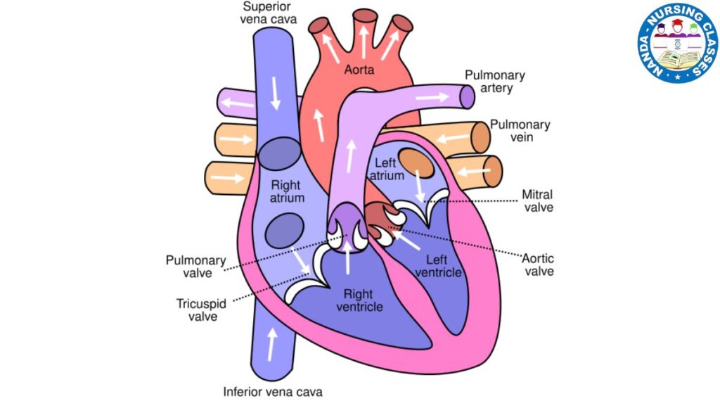
Anatomy of the Heart: You would learn that the heart has four main chambers (two atria and two ventricles), four valves that control blood flow, and its specific location within the chest (between the lungs, slightly to the left). You’d also identify the major blood vessels that bring blood to and from the heart.
-
Anatomy of a Bone: You’d discover that bones are composed of dense compact bone and porous spongy bone, contain blood vessels and nerves, and how they connect to other bones at joints.
What is Physiology?
Physiology is the study of the function of the body’s organs and systems. It focuses on how the different parts of the body work, how they control their activities, and how they integrate to maintain the overall function of the organism. If anatomy answers “What is it?”, physiology answers “How does it work?”
Examples:
-
Physiology of the Heart: You would understand how the heart muscles contract (the cardiac cycle), how electrical signals stimulate the heart to beat, and how it efficiently pumps blood throughout the body, distributing oxygen and nutrients.
-
Physiology of Respiration: You’d learn how the lungs take air in and out, how oxygen moves from the lungs into the blood, and how carbon dioxide moves from the blood into the lungs for exhalation (gas exchange).
Nursing Correlation: As a nurse, understanding both anatomy and physiology together is paramount. If you know the structure of the kidney (anatomy), you can then comprehend how the kidney filters blood and produces urine (physiology). When a patient is ill, recognizing which body part is structurally compromised and how that impacts its function is key to developing an effective care plan.
Levels of Organization: How Our Body is Built
The human body is a marvel of complexity, but its structure can be understood by breaking it down into a hierarchy of levels, each building upon the one below it. Imagine this as a pyramid, with the simplest parts at the base supporting every level above until they form the complete living you.
Here are the levels, from the smallest to the largest:
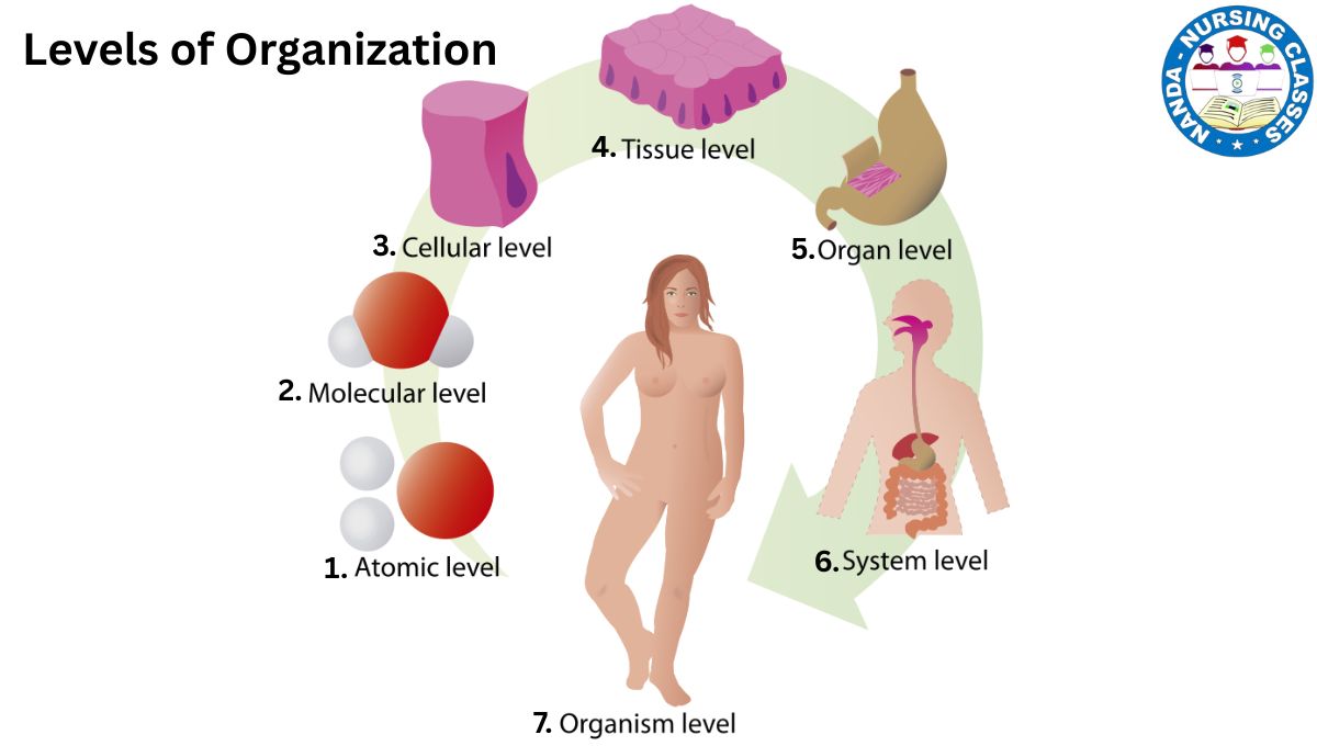
The Chemical Level: The Basic Building Blocks
- Atoms: This is the most basic level. It involves atoms, the smallest, indivisible particles of matter (like oxygen, carbon, hydrogen, nitrogen).
- Molecules: When these atoms bond together, they form molecules. Some important molecules for our body include water (H2O), proteins (which build muscles and enzymes), carbohydrates (for energy), lipids (fats), and nucleic acids (DNA and RNA, which carry our genetic information).
In simple terms: This is the level of ingredients before anything is built. These molecules are the fundamental basis for all structures and processes essential for life.
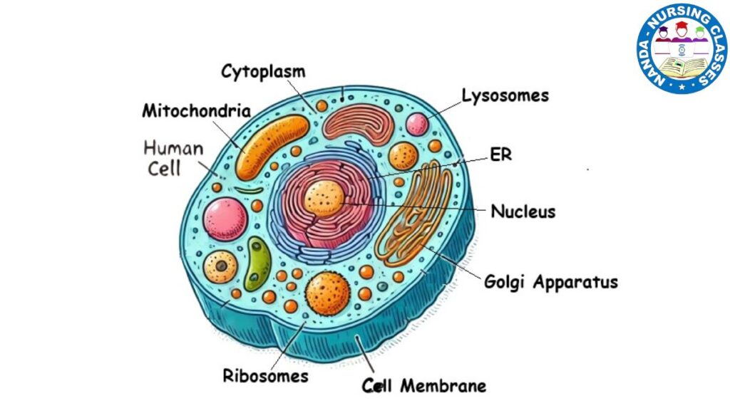
The Cellular Level: The Smallest Unit of Life
3. Cellular level: When molecules combine in specific ways, they form cells. The cell is the smallest unit of life—the basic building block of all living organisms. Your body contains trillions of cells, each a complex factory with a specific job. There are many different types, such as muscle cells, nerve cells, and blood cells.
Examples:
- Red Blood Cells: Specialized for carrying oxygen.
- Muscle Cells: Capable of contracting to produce movement.
- Nerve Cells (Neurons): Communicate by transmitting electrical signals.
- Skin Cells: Protect us from the external environment.
In simple terms: If the chemical level is ingredients, the cellular level is the individual bricks. A single brick isn’t a house, but you can’t build one without it.
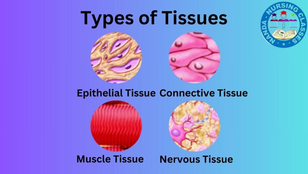
The Tissue Level: Cells Working Together
A tissue is a group of similar cells that work together to perform a specific function. It’s like cells forming a team where everyone has the same role. There are four primary types of tissue in the body:
- Epithelial Tissue: Covers body surfaces (like the outer layer of skin) and lines organs. It’s also involved in secretion and absorption.
- Connective Tissue: Connects, supports, and protects body organs. This includes bones, cartilage, blood, adipose (fat) tissue, and ligaments.
- Muscular Tissue: Responsible for generating movement, both internal and external. There are three types: skeletal (voluntary movement), smooth (found in organ walls, like the intestines), and cardiac (found only in the heart).
- Nervous Tissue: Communicates by transmitting electrical impulses between different parts of the body, allowing us to think, feel, and react. It’s found in the brain, spinal cord, and nerves.
In simple terms: Tissues are like the walls of the house, each made of many bricks (cells) and serving a different purpose (an outer wall vs. an inner wall).
Organ Level: A Functional Unit
An organ is a structure made up of two or more different types of tissues that work together to perform a complex, specific task. Different types of tissues come together to form a distinct organ, which performs a specific function. An organ typically contains two or more types of tissues.
Examples:
The Heart (an organ) is made of:
- Muscle tissue to pump blood.
- Nervous tissue to regulate the heartbeat.
- Connective tissue that holds it together and forms its valves.
- Epithelial tissue that lines its chambers.
-
Other examples include the stomach, liver, brain, and skin (which is also an organ), and others we will discuss later.
In simple terms: This is where the entire house is built. The wiring (nervous tissue), plumbing (connective tissue), and walls (muscle tissue) are all integrated to form a single, functional structure-the organ-with a major purpose, like pumping blood.
The Organ System Level: The Super-Teams
An organ system is a group of organs that work together to perform a major bodily function necessary for survival. A group of organs that work together to accomplish a major function for the body forms an organ system.
Our body has 11 major organ systems (e.g., Respiratory, Digestive, Cardiovascular, etc.).
Examples:
- Digestive System: Includes organs like the mouth, esophagus, stomach, intestines, liver, and pancreas. All these organs work together to break down food, absorb nutrients, and eliminate waste.
- Cardiovascular System: Composed of the heart, blood vessels, and blood, this system transports oxygen, nutrients, and hormones throughout the body.
In simple terms: Now, imagine a whole neighborhood. The house (organ) can’t function alone-it needs the power plant (nervous system) for electricity, the water treatment plant (urinary system) for clean water, and the grocery store (digestive system) for food. Organ systems are these interconnected networks that support each other.
Organismal Level: The Complete Being
This is the highest level of organization. It is the entire living being – all of the organ systems combined and working in harmony to maintain life. All organ systems work together to form a complete, living organism – a human body!
In simple terms: This is you. It is the sum of all other levels—chemical, cellular, tissue, organ, and organ system—functioning together in an integrated and self-sustaining way. It represents the complex, beautiful, and living whole.
In summary, The body is organized from simple to complex:
Atoms → Molecules → Organelles → Cells → Tissues → Organs → Organ Systems → The Organism.
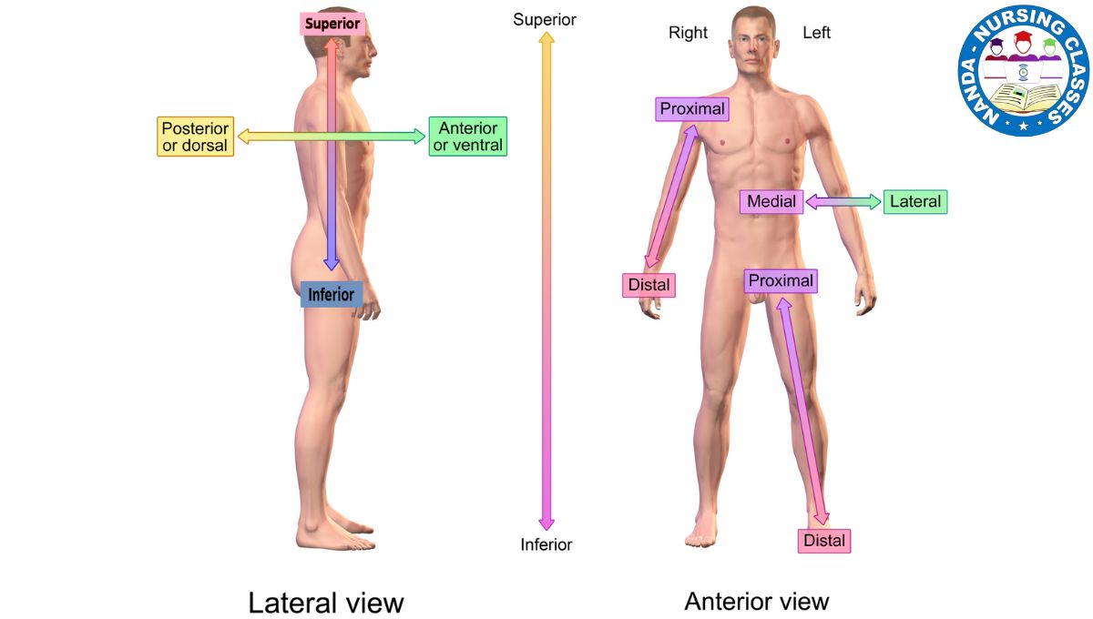
Anatomical terms
Anatomical terms are specialized vocabulary used in medicine, biology, and related fields to describe the locations, positions, movements, and relationships of different parts of the human (or animal) body. These terms help professionals communicate clearly and accurately, especially during procedures, studies, and diagnoses. Here’s an overview of key types of anatomical terms:
Directional Terms
These terms describe the relative position of body parts:
- Superior (Cephalic): Toward the head or upper part of a structure.
- Inferior (Caudal): Toward the lower part of the body.
- Anterior (Ventral): At or near the front of the body.
- Posterior (Dorsal): At or near the back of the body.
- Medial: Closer to the midline of the body.
- Lateral: Farther from the midline.
- Proximal: Closer to the point of attachment or origin.
- Distal: Farther from the point of attachment or origin.
- Superficial: Near the surface of the body.
- Deep: Farther from the body surface.
Body Movements
Movements describe changes in the position of body parts relative to one another:
- Flexion: Bending movement, decreasing the angle between bones.
- Extension: Straightening movement, increasing the angle between bones.
- Abduction: Movement away from the midline.
- Adduction: Movement toward the midline.
- Rotation: Movement around the longitudinal axis of a limb.
- Supination: Rotating the forearm so the palm faces up.
- Pronation: Rotating the forearm so the palm faces down.
Terms Related to Body Planes and Sections
Body planes are imaginary lines used to divide the body for anatomical study:
- Sagittal Plane: Divides the body into left and right sections.
- Transverse (Horizontal) Plane: Divides the body into upper (superior) and lower (inferior) parts.
- Frontal (Coronal) Plane: Divides the body into front (anterior) and back (posterior) sections.
- Oblique Plane: Cuts the body at an angle between the horizontal and vertical planes..
Anatomical Position and Body Symmetry
Anatomical Position
The standard reference position for describing locations and movements of body parts. In this position:
Bilateral Symmetry
The concept that the body can be divided into equal right and left halves along the midline, with each side mirroring the other.
Regional Names of Body Parts
To better describe specific areas, various regions of the body are named:
- Abdominal: The front part of the torso, below the diaphragm.
- Acromial: The shoulder area.
- Brachial: The arm.
- Carpal: The wrist.
- Cervical: The neck region.
- Femoral: The thigh.
- Lumbar: The lower back, between the ribs and pelvis.
- Thoracic: The chest region.
- Patellar: The front of the knee.
- Plantar: The sole of the foot.
These regional terms help precisely identify locations and guide medical practices.
Body Quadrants and Regions
The abdomen is divided for clinical and diagnostic purposes:
Nine Abdominal Regions:
-
- Right Hypochondriac
- Epigastric
- Left Hypochondriac
- Right Lumbar
- Umbilical
- Left Lumbar
- Right Inguinal (Iliac)
- Hypogastric
- Left Inguinal (Iliac)
Four Abdominopelvic Quadrants:
These quadrants and regions are used to locate organs and diagnose abdominal issues.
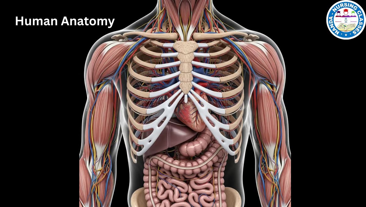
Systems and cavities of the human body
Systems of the Human Body
The human body is organized into multiple systems, each with specific functions and components:
- Integumentary System: Includes skin, hair, nails, sweat, and oil glands; provides protection and regulates body temperature.
- Skeletal System: Composed of bones and joints; supports and protects organs and allows for movement.
- Muscular System: Includes muscles; enables movement, posture, and heat production.
- Nervous System: Composed of the brain, spinal cord, and nerves; regulates body functions and processes sensory information.
- Cardiovascular System: Includes the heart and blood vessels; transports oxygen, nutrients, and waste.
- Lymphatic System: Composed of lymph nodes, lymph vessels, and lymph fluid; helps defend against infection.
- Respiratory System: Composed of lungs and airways; enables breathing and gas exchange.
- Digestive System: Includes organs from the mouth to the intestines; processes food, absorbs nutrients, and expels waste.
- Urinary System: Includes kidneys, bladder, and associated ducts; filters blood and eliminates waste.
- Reproductive System: Includes reproductive organs; responsible for producing gametes and supporting reproduction.
Body Cavities
Body cavities are spaces within the body that house and protect vital organs, allowing them to expand, move, and function properly. The two main types of body cavities are the dorsal cavity and the ventral cavity.
- Dorsal Cavity
The dorsal cavity is located along the back (posterior) of the body and is divided into two main cavities:
- Cranial Cavity: Located within the skull, this cavity houses and protects the brain.
- Spinal (Vertebral) Cavity: Runs along the length of the spinal column and contains the spinal cord, which is protected by the vertebrae.
Together, the cranial and spinal cavities form the dorsal cavity, which primarily protects the central nervous system.
- Ventral Cavity
The ventral cavity is located along the front (anterior) of the body and is divided into two major cavities, which are separated by the diaphragm (a large, dome-shaped muscle used in breathing):
- Thoracic Cavity: The upper part of the ventral cavity, located above the diaphragm, which contains:
- Pleural Cavities: Two cavities that each contain a lung.
- Mediastinum: The central area between the lungs, containing the heart, trachea, esophagus, and major blood vessels.
- Pericardial Cavity: A cavity within the mediastinum that encloses the heart.
- Abdominopelvic Cavity: The lower part of the ventral cavity, located below the diaphragm, which is further divided into:
- Abdominal Cavity: The upper portion, containing organs such as the stomach, liver, intestines, pancreas, spleen, and kidneys.
- Pelvic Cavity: The lower portion, which houses the bladder, reproductive organs, and parts of the intestines.
Functions of Body Cavities
- Protection: Cavities shield delicate organs like the brain, heart, and lungs from physical damage.
- Organ Expansion: Cavities provide space for organs to expand, such as the lungs during breathing and the stomach after eating.
- Isolation of Functions: By housing specific organs, cavities help contain infection and inflammation within particular areas of the body, preventing them from spreading.
Conclusion
Anatomical terms serve as a standardized language for describing the human body, allowing clear communication across medical, biological, and scientific fields. By understanding terms for direction, movement, body planes, and regions, professionals can accurately describe the locations, functions, and movements of various body parts. This precision is vital for diagnoses, medical procedures, and scientific research, minimizing ambiguity and ensuring that everyone involved understands the body in the same way.
Anatomy and Physiology learn in Hindi
FAQs
Q1: Why are anatomical terms important?
Anatomical terms are essential for clear and precise communication in medicine and biology. They help professionals accurately describe locations, movements, and relationships between body parts, which is crucial for diagnoses, surgeries, and research.
Q2: What is the difference between anterior and posterior?
Anterior (or ventral) refers to the front of the body, while posterior (or dorsal) refers to the back. For example, the chest is anterior to the spine, and the spine is posterior to the chest.
Q3: What is the sagittal plane?
The sagittal plane is an imaginary line that divides the body into left and right parts. It is one of the main anatomical planes used to describe positions and movements in the body.
Q4: How do flexion and extension differ?
Flexion decreases the angle between two body parts (e.g., bending the elbow), while extension increases this angle (e.g., straightening the elbow). These terms describe common movements in joints like the knees and elbows.
Q5: What do terms like “medial” and “lateral” mean?
Medial means closer to the midline of the body, while lateral means farther from the midline. For example, the nose is medial to the eyes, and the ears are lateral to the eyes.
Q6: Can anatomical terms apply to animals as well as humans?
Yes, anatomical terms can be used to describe the body structures of both humans and animals, especially in veterinary medicine and comparative anatomy, though some species-specific terms may differ.
Please comment on any terminology that you want to Know.


Hello!
Good cheer to all on this beautiful day!!!!!
Good luck 🙂